What is Craniopharyngioma?
Craniopharyngiomas are benign tumors. These occur at the base of the brain, right above the pituitary gland. The term has been derived from three different terms:
Cranio + pharyng + oma
1. Cranio – head
2. Pharyng – throat
3. Oma – condition
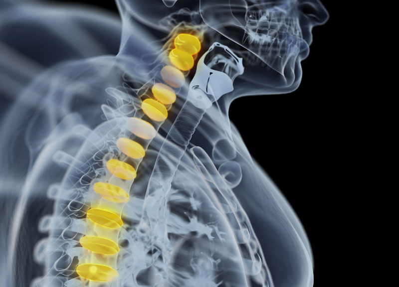
Craniopharyngiomas usually involves the pituitary stalk. It actually serves as the link between the pituitary gland and the brain.
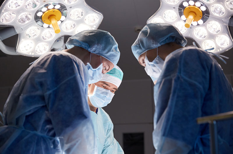
Carniopharyngioma – Expert Solution and Care at Premier Brain & Spine
Our Neurosurgeons at Premier Brain & Spine offer comprehensive management to treat carniopharyngiomas. We have many years of experience in diagnosing, treating and managing varied types of pituitary conditions. We use the most advanced diagnostic tools to properly diagnose these tumors.
Understanding Craniopharyngioma
Both adults and children may develop craniopharyngiomas. Most children are diagnosed with the condition between 5-10 years of age. The condition is rare. Craniopharyngiomas have a tendency to grow large, sometimes bigger than a golf ball. The patient may not experience any symptoms in the beginning.
Craniopharyngiomas – The Types
Adamantinomatous Craniopharyngioma – Under the microscope, these cells look very similar to the cells that produce tooth enamel. This type commonly calcifies, which is visible on computed tomography (CT) scanning and helpful in making the diagnosis.
Papillary Craniopharyngioma – This type rarely calcifies.
Symptoms of Craniopharyngioma
The symptoms produced by a craniopharyngioma will vary depending on the following factors:
– Exact location of tumor
– Age of patient
– The specific hormone involved
Common symptoms include:
- Changes in personality
- Confusion
- Headache
- Vomiting
Pituitary insufficiency is another symptom seriously considered by doctors. This occurs due to some degree as craniopharyngiomas may develop in the area of the pituitary stalk. This has an adverse effect on the overall function of the pituitary gland.
Children affected by hormone deficiency will show visible symptoms such as failure in growth and delayed puberty.
Other Symptoms:
Diabetes insipidus – This results due to the absence of antidiuretic hormone (ADH), a posterior pituitary hormone. The condition is further determined by symptoms such as excessive thirst and excessive urination
Adrenal insufficiency – This occurs due to reduction in ACTH production and cortisol reduction. This may be fatal under severe cases. The condition is characterized by symptoms such as fatigue, low blood pressure and electrolyte abnormalities.
Growth hormone (GH) insufficiency – This results due to the reduction in growth hormone (GH) production. The condition is determined by symptoms including stunted growth and delayed puberty in children, general fatigue and loss of muscle mass and tone in adults.
Hypothyroidism – The condition results from a reduction in TSH production. It is determined by certain visible symptoms such as loss of appetite, fatigue, weight gain, and decreased mental function.
Prolactin Production Reduction – The condition is rare and occurs with severe pituitary insufficiency.
Large pituitary tumors – The “stalk effect” paradoxically elevates blood prolactin levels due to the compression of the pituitary stalk interfering with control of brain over prolactin production.
Premenopausal Women – The elevated prolactin may result in a reduction or loss of menstrual periods and/or production of breast milk (galactorrhea).
Stalk Effect – This slightly elevates prolactin levels as opposed to prolactinomas wherein the level of prolactin is very high.
Reduction of Sex Hormones – The body experiences reduction of luteinizing hormone (LH) and follicle-stimulating hormone (FSH). Men suffer from low testosterone level leading to decreased sexual drive and impotence. In some cases, men also experience loss of body and facial hair. Women suffer from infertility.
Vision Loss and Craniopharyngiomas
This occurs when the tumor involves the optic tracts, chiasm, or nerves.
Symptoms occurring due to the affected hypothalamus in a region at the base of the brain may include:
- Diabetes insipidus
- Obesity
- Increased drowsiness / excess sleepiness
- Abnormalities associated with temperature regulation
Diagnosis for Craniopharyngioma
The doctor conducts a physical examination in detail. He/she will ask about symptoms and medical history. Diagnostic procedures will include:
– Testing of pituitary function
– Imaging scans
Imaging Scans for Craniopharyngioma
MRI Scan – The doctor suggest a high-resolution magnetic resonance imaging (MRI) scan. The test is considered very valuable as it helps the neuroradiologist to view the tumor closely and from different angles.
3T (Tesla) MRI Scanner – A powerful 3T (Tesla) MRI scanner is used by doctors in some cases. This helps them define the exact location of critical brain structures that may be affected by the tumor.
Pituitary Functioning Test – The testing is very important for patients with a Craniopharyngioma. Endocrinologist holding specialization in pituitary tumors will help assess the results in detail.
CT Scan – Doctors prefer a computed tomography (CT) scan. It is also a powerful diagnostic tool to detect calcification of the tumor
Treatment Options for Craniopharyngioma
A medical team comprising of specialists from diverse disciplines will work together to manage patient’s treatment. The program at Premier Brain & Spine has experts in each of the specialities. All of these professionals work closely together so as to provide you with the most comprehensive, state-of-the-art surgical treatments. Nonsurgical treatments are also provided according to the condition of the patient.
Treatment options include:
– Surgery
– Medication
– Radiation Therapy
Surgery for Craniopharyngioma
Endoscopic Endonasal Surgery
When diagnosed with Craniopharyngioma, the patient may require surgery:
– Establish or confirm the diagnosis.This is done via removal of a sample of tumor tissue for detailed analysis.
– Decompress optic chiasm (part of the brain where the optic nerves cross partially) to improve vision.
– Remove as much of the tumor as possible without producing potential risks.
There are many different surgical approaches to Craniopharyngioma tumors. The medical team at Premier Brain & Spine will decide on a specific approach to use based on the following factors:
- Location of the tumor with respect to key brain structures
- Whether the tumor is cystic (contains fluid-filled cavities)
- Size of the tumor
- The extent to which the tumor invaded surrounding structures, especially the hypothalamus
- Overall health of patient
- Age of patient
The Goal of Surgery:
It differs for both children and adults:
Children – The surgeon will try his best to remove the entire tumor and as safely as possible. He may also recommend a minimally invasive endoscopic endonasal approach. This is important because it can cause a very less amount of hypothalamic injury. This means the patient will have no visible scar post surgery.
Adults – Doctors usually often use a more conservative approach with adults. It is aimed at establishing a diagnosis and decompressing the optic nerves. However, this is not done to fully remove the tumor. Since the tumor involves the pituitary stalk, any attempt to remove the entire tumor leads to complete pituitary insufficiency.
Surgical Approaches for Craniopharyngioma
The two basic approaches include:
– Endonasal
– Craniotomy
Endoscopic Endonasal Surgery
This kind of surgical approach is precisely a minimally invasive approach. Here, the surgeon will use natural nasal passageway of the patient. The process will not require a head incision. The neurosurgeons at Premier Brain & Spine are equipped with knowledge, with advanced training and extensive experience requisite for performing this complicated procedure.
The endoscopic technique is known to be very effective in removing the tumor without producing any risks and minimizing hospitalization time and discomfort at the same time.
The Drawback – Not every Craniopharyngioma tumor will be removed through endonasal endoscopic approach.
Do you want to find out if you are a potential candidate for an endoscopic approach? We will suggest you to arrange a meeting / consultation session with our medical experts.
Open Cranitomy Surgery for Craniopharyngiomas
The Process
The surgeon will do the following during an open craniotomy:
- Create an incision into patient’s scalp
- Remove a piece of skull temporarily
- Get very high magnification of the affected area through surgical microscope
- Elevates the base of the brain’s frontal lobe (above the eye) very gently to gain access to and remove the tumor
- The surgeon will use a minimally invasive eyebrow incision keyhole approach
Complications of Surgery
The following complications may occur with surgery in children:
- A complete resection of craniopharyngioma tumor may sometimes results in complete pituitary insufficiency. This will require patients undergo hormone replacement therapy, lifelong.
- Hypothalamus damage may lead several issues such as severe obesity.
The following complications may occur with surgery in both adults and children:
- Diabetes insipidus
- Optical nerves causing blindness
- Endonasal approach used by the surgeon may result in cerebrospinal fluid (CSF) leakage through the nose. This demands repeat surgery.
Craniopharyngiomas and Radiation Therapy
Sometimes, doctors may recommend radiation therapy immediately after surgery. In this case, a stereotactic radiosurgery is done. It is a a technique that makes use of highly focused radiation dosage delivered directly to the tumor.
The process is safe as the radiation beam is sculpted in a fashion to target only the tumor. This way, the surrounding brain structures are exposed to only a fraction of the radiation and remain potentially unharmed.
At the Premier Brain & Spine, we use the advanced Radiosurgery delivery system that allows us to treat all kinds of irregularly shaped tumors, even those close to critical brain structures.
The Drawback
The major drawback of radiation treatment is that it may result in pituitary failure in the long run. This occurs many years after treatment. Pituitary failure demands complete hormone replacement.
Pituitary Hormonal Dysfunction Treatment for Craniopharyngioma
The dysfunction is quite common post Craniopharyngioma surgery and/or radiation therapy. Issues with hypothalamic function may be quite a challenge for doctors to treat. The patient usually requires very careful monitoring combined with follow-up visits with an endocrinologist.