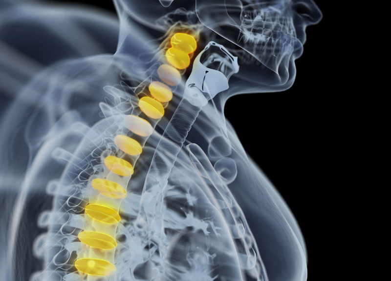
What is a Brain Aneurysm?
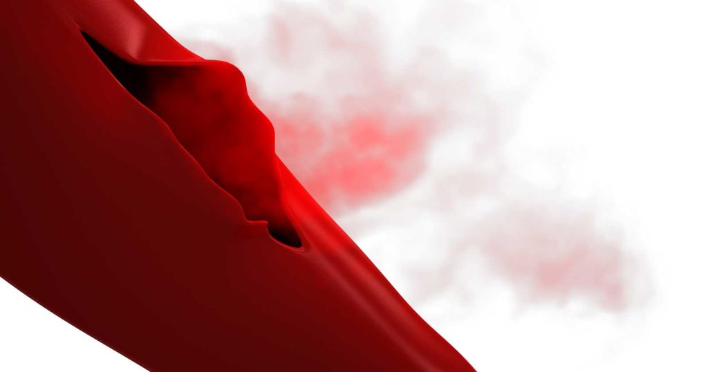
An aneurysm can be explained as an abnormal, weak spot on a blood vessel resulting in ballooning or bulging on the outward side of the artery wall. These spots can include all blood vessel walls (fusiform aneurysm) to create a sac from a single wall (secular). It may also separate vessel walls (dissecting). Any vessel in the body can be affected by an aneurysm. However, it is only when the weak spot reaches the vessel in head that leads to a serious medical condition. Referred to as a hemorrhagic stroke, this condition occurs when weak spots in the head, resulting in brain damage and ultimately death.
Most brain aneurysms are discovered when they rupture, which results in a subarachnoid hemorrhage due to bleeding into the brain or the subarachnoid space (the space surrounding the brain). This kind of hemorrhage may result in a hemorrhagic stroke, brain damage and ultimately death.
Major treatment goals of a ruptured aneurysm include:
- Stop bleeding
- Prevent any potential permanent damage to the brain
- Minimize the risk of recurrence
Symptoms of Ruptured Cerebral Aneurysm
Subarachnoid hemorrhage or ruptured aneurysm results in severe (very severe) headache. Most people define it as the worst headache experience of their life. Here is the list of other symptoms experienced by patients:
- Nausea
- Vomiting
- Stiff neck
- Neck pain
- Blurred vision
- Loss of sensation
- Double vision
- Pain above and behind the eye
- Dilated pupils
- Sensitivity to light
Symptoms of Unruptured Cerebral Aneurysm
Most aneurysms (the smaller ones) are asymptomatic. Sometimes, large aneurysms may result in the following symptoms. These symptoms are associated with pressure on the brain or adjacent nerves:
- Peripheral vision deficits
- Complications with speech
- Problems with thinking
- Issues with processing
- Perceptual problems
- Sudden changes in behaviour
- Loss of balance
- Loss of coordination
- Decreased concentration
- Fatigue
- Short-term memory difficulty
Since brain aneurysms symptoms may also be associated with other medical conditions, doctors prefer the use of diagnostic Neuroradiology for identifying ruptured and unruptured brain aneurysms.
Diagnosis
Aneurysms are known to rupture at about 1-2 per cent on a yearly basis. However, these tend to vary in size and location. Unfortunately, the presence of most aneurysms is an indication of rupture. These results in symptoms such as changes in vision, pain above and behind the eye, paralysis of nerve, pain in neck, localized headache, nausea and vomiting, etc.
CT (computed tomography) and MRI (magnetic resonance imaging) help with the determination of a large number of aneurysms way before they are ruptured. Both are noninvasive methods use by a radiologist to have a close look at the blood vessels in the head.
MR does not involve radiation or contrast risks. On the other hand, a CT produces better resolution and helps with an efficient operative planning. CT scan of the head and a CT angiogram will help diagnose aneurysm and subarachnoid hemorrhage.
Although CT and MR are capable of showing aneurysms, most patients require a cerebral angiogram which ensures definitive diagnosis. The process also helps determine the best treatment process. An angiogram is an invasive procedure. It involves Neuro-Interventional surgeon inserting a catheter (flexible tube) through an artery over the hip which is guided to the brain vessels. Thereafter, a liquid dye / contrast agent is injected into the vessel. Fluoroscope is used to take pictures. An angiogram is known to provide with the highest detailed pictures for exact location, size, and shape of the aneurysm. The information is used by doctor to plan the best treatment option for each patient.
Treatment Options
Not all aneurysms require treatment. A medical practitioner will decide on which one needs to be given treatment. According to medical experts, the following two methods are very effective in treating brain aneurysm:
1. Open Surgery
2. Endovascular Techniques
The best treatment option depends varied factors such as the aneurysm type (ruptured / unruptured), size, location and shape. It is best to allow your doctor to decide on the ideal treatment option.
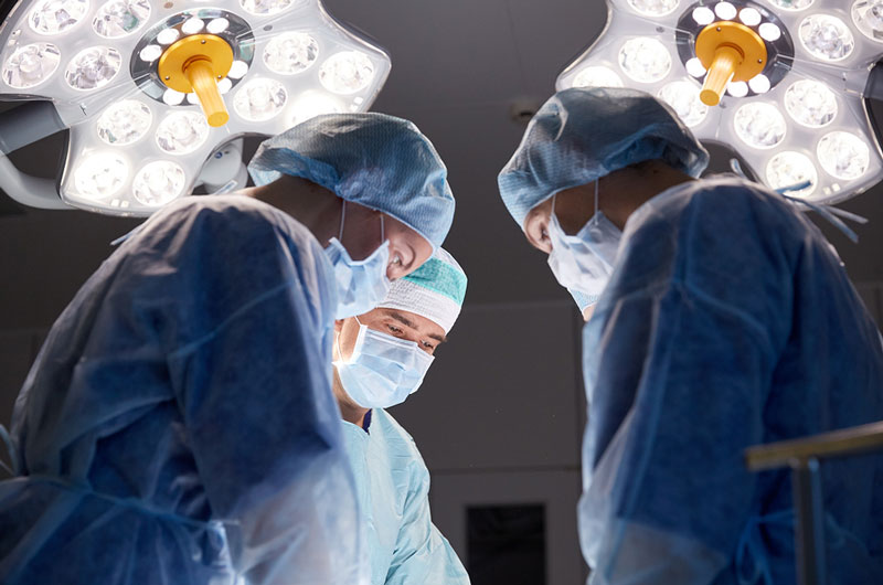
Open Surgery – “Clipping”
Performed by a neurosurgeon, the process involves making an incision in the skin over the head. The surgeon also makes an opening in the bone. A clip is placed across aneurysm via dissecting through the spaces in the brain. The clip is placed right at the point where it arises from the blood vessel. The process prevents flow of blood flow from entering the aneurysm.
Post surgery, patients is expected to spend 2-3 nights in the hospital. Thereafter, they are allowed to go home. The Doctor allows only light restricted activities for around 1-2 months.
With advancement in the techniques, neurosurgeons can now perform open surgery as mini craniotomies (eye brow incisions) in order to clip an aneurysm. A tiny incision can also be made over the eyebrow of the patient as per requirement. Thereafter, the surgeon will make a small window measuring 2 inches in the bone over the eye. A small clip is placed through this incision across aneurysm opening.
Endovascular “Coiling”
The treatment is performed by a neurointerventional surgeon. The professional may be a neurologist or neuroradiologist, with additional training. This treatment is performed as an extended process of angiogram and best recommended for ruptured aneurysm.
The surgeon will insert a catheter into a vessel over the hip. Other catheters are navigated through the blood vessels travelling to other vessels in the brain and further into the aneurysm. Thereafter, the coils are packed into the aneurysm to the point where it arises from the blood vessel. This prevents the flow of blood from entering the aneurysm.
The process may also need some additional devices, such as a balloon or a stent to keep the coils in place within the aneurysm. Coiling that involves the use of stent require permanent placing of a stent inside vessel next to the aneurysm. This done with an aim to provide support to keep coils within aneurysm sac.
The Balloon remodeling process involves placement of a removable balloon on a temporary basis next to the aneurysm. The coils are positioned inside the aneurysm.
After the treatment, most patients are allowed to go home the next day. They can also get back to their normal activities the following day. With significant advances in endovascular techniques, surgeons are using new flow that diverts immobilization devices. These devices are very similar to a stint in a manner that they can be placed inside the main vessel located next to an aneurysm.
The devices are helpful in diverting flow away from the aneurysm. These ensure healing of the vessel wall and disappearing of the aneurysm with time. Doctors can treat intreatable aneurysms or the ones labelled as ‘high risk’ by other methods.
A number of new devices are available these days such as newer ones. These are very easy and safe to deliver.